Non-healing leg wounds represent one of the most challenging clinical problems in modern healthcare, affecting millions of people worldwide and imposing significant burden on both patients and healthcare systems. These chronic wounds, which fail to progress through the normal healing stages within an expected timeframe, often signal underlying pathophysiological processes that require comprehensive evaluation and specialised treatment approaches. The complexity of lower extremity wound healing involves intricate interactions between vascular supply, cellular mechanisms, inflammatory responses, and systemic factors that can become disrupted in various disease states.
Understanding why a simple leg cut transforms into a persistent, non-healing ulcer requires examining the fundamental mechanisms that govern tissue repair and regeneration. When these processes become compromised, what should be a straightforward healing journey can extend for months or even years, leading to substantial morbidity and healthcare costs. The multifactorial nature of chronic leg wounds demands a thorough understanding of both local and systemic contributing factors to develop effective treatment strategies.
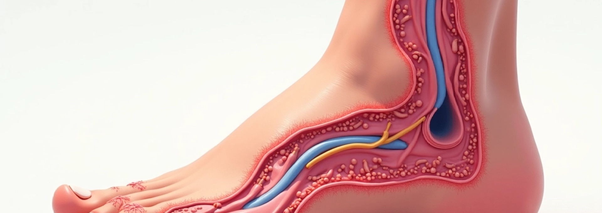
Pathophysiology of Non-Healing leg wounds: underlying mechanisms
The transformation of an acute wound into a chronic, non-healing lesion involves complex disruptions in the normal wound healing cascade. Understanding these pathophysiological mechanisms is crucial for developing targeted therapeutic interventions and improving patient outcomes. Chronic wounds typically become trapped in a prolonged inflammatory phase, preventing progression through the normal stages of hemostasis, inflammation, proliferation, and remodelling.
Impaired angiogenesis and capillary dysfunction in chronic wounds
Angiogenesis, the formation of new blood vessels, represents a critical component of successful wound healing. In chronic leg wounds, this process becomes significantly impaired due to various factors including hypoxia, elevated protease activity, and reduced growth factor availability. The inability to establish adequate vascularisation perpetuates tissue hypoxia and nutrient deficiency, creating a self-perpetuating cycle of poor healing.
Endothelial cell dysfunction in chronic wounds manifests through reduced nitric oxide production, increased vascular permeability, and impaired cellular migration. These alterations compromise the formation of stable, functional capillary networks essential for delivering oxygen and nutrients to the wound bed. Research indicates that chronic wounds exhibit significantly reduced capillary density compared to acute wounds, with measurements showing up to 40% fewer functional vessels in the wound periphery.
Dysregulated inflammatory response and cytokine imbalance
The inflammatory phase of wound healing normally resolves within days to weeks, transitioning to the proliferative phase. However, chronic leg wounds demonstrate persistent inflammation characterised by elevated levels of pro-inflammatory cytokines including interleukin-1β, tumour necrosis factor-α, and interleukin-6. This sustained inflammatory state prevents the wound from progressing to subsequent healing phases.
Macrophage polarisation plays a crucial role in this dysfunction, with chronic wounds showing predominance of M1 (classically activated) macrophages rather than the M2 (alternatively activated) phenotype required for healing progression. The imbalance between matrix metalloproteinases (MMPs) and their inhibitors further contributes to tissue destruction, with chronic wounds showing up to 30-fold elevation in MMP-2 and MMP-9 activity compared to healing wounds.
Extracellular matrix degradation and collagen synthesis disruption
The extracellular matrix provides essential scaffolding for cellular migration, proliferation, and tissue reconstruction. In chronic leg wounds, excessive protease activity leads to continuous degradation of newly synthesised collagen and other matrix proteins. This creates an environment where tissue destruction outpaces tissue formation, preventing wound closure and epithelialisation.
Fibroblast dysfunction represents another critical factor, with cells in chronic wounds showing reduced proliferative capacity, altered gene expression profiles, and decreased collagen production. Studies demonstrate that fibroblasts from chronic wounds exhibit senescence-like characteristics , with shortened telomeres and reduced response to growth factor stimulation. This cellular dysfunction contributes to the formation of weak, poorly organised tissue that remains vulnerable to reinjury.
Bacterial biofilm formation and antimicrobial resistance patterns
Biofilm formation represents a sophisticated bacterial survival strategy that significantly complicates chronic wound management. These organised microbial communities, encased in self-produced polymeric matrices, demonstrate remarkable resistance to antimicrobial therapy and host immune responses. Research indicates that over 60% of chronic wounds harbour bacterial biofilms, compared to less than 6% of acute wounds.
The polymicrobial nature of chronic wound biofilms creates complex interactions between different bacterial species, often including Staphylococcus aureus, Pseudomonas aeruginosa, and anaerobic organisms. These communities can persist despite appropriate antibiotic therapy, with biofilm-associated bacteria showing up to 1,000-fold increased resistance to antimicrobials. The presence of biofilms also perpetuates inflammation and interferes with normal cellular processes required for healing.
Vascular insufficiency: arterial and venous compromise assessment
Vascular compromise represents the most common underlying cause of non-healing leg wounds, encompassing both arterial insufficiency and venous dysfunction. The lower extremities face unique challenges due to their dependent position and the requirement for blood to flow against gravity during venous return. Comprehensive vascular assessment forms the cornerstone of chronic wound evaluation, as addressing underlying vascular pathology is essential for achieving successful healing outcomes.
Peripheral arterial disease and Ankle-Brachial index evaluation
Peripheral arterial disease (PAD) affects approximately 8-10% of adults over 65 years, with significantly higher prevalence among diabetic patients and smokers. The condition results from atherosclerotic narrowing or occlusion of peripheral arteries, leading to reduced blood flow and tissue hypoxia. PAD-related wounds typically present with characteristic features including well-demarcated borders, minimal exudate, and severe pain that worsens with elevation.
The ankle-brachial index (ABI) serves as the primary screening tool for PAD, comparing systolic blood pressure measurements between the ankle and arm. Normal ABI values range from 1.0 to 1.3, while values below 0.9 indicate arterial insufficiency. However, patients with diabetes or chronic kidney disease may demonstrate falsely elevated ABI readings due to arterial calcification, necessitating additional testing such as toe-brachial index or transcutaneous oxygen measurements. Critical limb ischaemia , defined by ABI values below 0.4 or toe pressures below 30 mmHg, represents a medical emergency requiring urgent vascular intervention to prevent amputation.
Chronic venous insufficiency and duplex ultrasound findings
Chronic venous insufficiency (CVI) affects approximately 25% of adults and represents the leading cause of leg ulceration in developed countries. The condition results from valvular incompetence, venous obstruction, or calf muscle pump dysfunction, leading to venous hypertension and subsequent tissue damage. CVI-related ulcers typically develop in the gaiter area, particularly around the medial malleolus, and are often accompanied by oedema, skin changes, and varicose veins.
Duplex ultrasound assessment provides crucial information about venous anatomy, valve function, and flow dynamics. The examination evaluates both superficial and deep venous systems, identifying reflux duration, obstruction patterns, and perforator vein incompetence. Venous reflux lasting more than 0.5 seconds in superficial veins or 1.0 second in deep veins indicates significant valvular dysfunction. The CEAP classification system (Clinical, Etiology, Anatomy, Pathophysiology) provides standardised assessment of CVI severity, helping guide treatment decisions and predict healing outcomes.
Mixed arteriovenous pathology and haemodynamic assessment
Mixed arteriovenous disease presents particular diagnostic and therapeutic challenges, as patients may simultaneously exhibit features of both arterial insufficiency and venous hypertension. This combination occurs in approximately 15-25% of leg ulcer patients and requires careful balance between improving arterial perfusion and managing venous congestion. The presence of mixed pathology significantly complicates treatment decisions, particularly regarding compression therapy, which may be contraindicated or require modification in patients with arterial compromise.
Haemodynamic assessment in mixed disease requires comprehensive evaluation using multiple modalities including ABI, venous duplex scanning, and advanced techniques such as air plethysmography or photoplethysmography. These investigations help quantify the relative contributions of arterial and venous pathology, guiding individualised treatment approaches. Patients with mixed disease often experience prolonged healing times and higher recurrence rates compared to those with isolated vascular pathology.
Microvascular disease in diabetic patients and transcutaneous oxygen measurement
Diabetic microvascular disease represents a distinct pathophysiological entity characterised by basement membrane thickening, endothelial dysfunction, and impaired autoregulation. Unlike macrovascular disease, diabetic microangiopathy affects the smallest vessels, leading to tissue hypoxia despite apparently adequate macrovascular flow. This condition particularly affects the feet and lower legs, contributing to the high incidence of diabetic foot ulcers and their notorious resistance to healing.
Transcutaneous oxygen pressure (TcPO2) measurement provides valuable assessment of tissue oxygenation in diabetic patients. Normal TcPO2 values exceed 40 mmHg, while values below 20 mmHg indicate severe hypoxia unlikely to support healing without intervention. The test can also evaluate response to oxygen supplementation, helping predict outcomes with hyperbaric oxygen therapy. Research demonstrates that diabetic patients with TcPO2 values above 25 mmHg show significantly better healing rates compared to those with lower measurements, making this investigation crucial for treatment planning.
Systemic conditions contributing to delayed wound healing
Numerous systemic conditions can significantly impair wound healing through various mechanisms including altered immune function, impaired protein synthesis, medication effects, and metabolic disturbances. Diabetes mellitus stands as the most significant systemic contributor to non-healing leg wounds, affecting approximately 463 million adults worldwide and substantially increasing ulceration risk. The multifactorial nature of diabetic wound healing impairment involves hyperglycaemia-induced protein glycation, advanced glycation end product formation, and compromised immune cell function.
Chronic kidney disease creates a complex milieu of healing impairment through uremic toxin accumulation, chronic inflammation, mineral bone disorders, and anaemia. Patients with end-stage renal disease demonstrate significantly prolonged healing times, with studies showing up to 50% longer healing duration compared to patients with normal kidney function. The uraemic environment also promotes calciphylaxis, a devastating condition characterised by vascular calcification and subsequent tissue necrosis.
Autoimmune conditions such as rheumatoid arthritis, systemic lupus erythematosus, and vasculitis can contribute to wound healing impairment through direct vascular involvement, chronic corticosteroid use, and disease-modifying antirheumatic drug effects. Vasculitic ulcers often present with characteristic features including irregular borders, surrounding purpura, and severe pain disproportionate to wound size. These conditions require careful coordination between wound care specialists and rheumatologists to balance disease control with healing promotion.
Malnutrition represents an often-overlooked contributor to delayed healing, with protein-energy malnutrition affecting up to 20% of elderly patients with chronic wounds. Inadequate protein intake impairs collagen synthesis, immune function, and cellular proliferation, while deficiencies in specific nutrients including vitamin C, zinc, and arginine further compromise healing capacity. Comprehensive nutritional assessment and intervention form essential components of chronic wound management, particularly in elderly or institutionalised patients.
The presence of multiple comorbidities creates a synergistic effect on healing impairment, with patients having three or more systemic conditions showing exponentially increased healing times and complication rates.
Advanced diagnostic approaches for chronic leg ulcers
Contemporary chronic wound assessment extends far beyond visual inspection, incorporating sophisticated diagnostic techniques that provide molecular-level insights into wound pathophysiology. Advanced diagnostics enable precise characterisation of healing barriers, guide targeted therapy selection, and monitor treatment response objectively. The integration of these technologies represents a paradigm shift from empirical treatment approaches toward precision wound care.
Bacterial diagnostics have evolved significantly beyond traditional culture methods, which often fail to detect biofilm-associated organisms and may require several days for results. Molecular diagnostic techniques including polymerase chain reaction (PCR) and next-generation sequencing provide rapid, comprehensive microbial profiling that identifies fastidious organisms, quantifies bacterial burden, and detects antimicrobial resistance genes. These techniques reveal that chronic wounds harbour significantly more diverse microbial communities than previously recognised, with implications for targeted antimicrobial therapy.
Wound fluid biomarker analysis represents an emerging diagnostic frontier, utilising proteomic and genomic approaches to characterise the molecular wound environment. Elevated levels of inflammatory cytokines, matrix metalloproteinases, and bacterial endotoxins can indicate healing impairment before clinical signs become apparent. Point-of-care devices now enable rapid measurement of wound pH, bacterial fluorescence, and tissue oxygenation, providing immediate feedback to guide treatment decisions.
Advanced imaging modalities including optical coherence tomography, hyperspectral imaging, and thermal imaging provide non-invasive assessment of wound characteristics, perfusion patterns, and healing progression. These technologies can detect subclinical changes in tissue viability, identify areas at risk for breakdown, and monitor treatment response with unprecedented precision. Hyperspectral imaging can detect tissue oxygenation changes up to 48 hours before clinical signs of healing impairment become apparent, enabling proactive intervention.
Tissue biopsy remains crucial for excluding malignancy, particularly in wounds that fail to respond to appropriate therapy or demonstrate atypical characteristics. Studies indicate that up to 3% of chronic leg ulcers harbour underlying malignancy, most commonly squamous cell carcinoma arising in long-standing wounds (Marjolin’s ulcer). Histological examination also provides information about tissue architecture, inflammatory patterns, and the presence of specific pathogens that may guide therapeutic decisions.
Evidence-based treatment modalities and wound care technologies
Contemporary chronic wound management has evolved into a sophisticated discipline incorporating evidence-based approaches, advanced technologies, and personalised treatment strategies. The foundation of effective treatment remains addressing underlying pathophysiology while optimising local wound conditions to promote healing. This multifaceted approach requires careful consideration of wound characteristics, patient factors, and available resources to develop individualised treatment plans.
Negative pressure wound therapy and VAC system applications
Negative pressure wound therapy (NPWT) has revolutionised chronic wound management since its introduction, providing controlled subatmospheric pressure that promotes healing through multiple mechanisms. The therapy enhances local blood flow, reduces oedema, removes excess exudate, and mechanically stimulates cellular proliferation. Clinical trials demonstrate that NPWT can reduce healing time by 30-50% compared to conventional dressings in appropriate wound types.
The mechanism of action involves applying controlled negative pressure (typically -75 to -125 mmHg) through a sealed foam or gauze interface, creating mechanical forces that stimulate angiogenesis and granulation tissue formation. The therapy also reduces bacterial burden by removing contaminated exudate and creating an environment unfavourable for pathogen proliferation. Modern NPWT systems incorporate advanced features including programmable pressure cycles, instillation capabilities for delivering topical therapies, and portable devices that maintain patient mobility.
Patient selection for NPWT requires careful consideration of wound characteristics, vascular status, and contraindications. The therapy demonstrates particular efficacy in deep wounds with adequate vascular supply, post-surgical dehiscence, and traumatic injuries. However, NPWT is contraindicated in the presence of untreated osteomyelitis, malignancy within the wound bed, or exposed blood vessels. Proper patient education and follow-up protocols are essential for maximising therapy benefits and preventing complications.
Bioengineered skin substitutes and cellular therapy options
Bioengineered skin substitutes represent a major advancement in chronic wound treatment, providing biological scaffolds that support cellular migration, proliferation, and tissue reconstruction. These products range from acellular dermal matrices to living cellular constructs containing fibroblasts, keratinocytes, or stem cells. The selection of appropriate skin substitutes depends on wound characteristics, healing phase, and specific clinical requirements.
Cellular therapy approaches include autologous and allogeneic options, with living skin equivalents demonstrating particular efficacy in venous and diabetic ulcers. Clinical studies show that patients treated with cellular therapies achieve
significantly higher healing rates, with complete closure achieved in 50-70% of patients within 12 weeks compared to 20-30% with standard care alone.
Stem cell therapy represents the cutting edge of regenerative wound care, utilising mesenchymal stem cells, adipose-derived stem cells, or platelet-rich plasma to enhance healing capacity. These therapies work by releasing growth factors, modulating inflammation, and promoting angiogenesis. Autologous treatments using the patient’s own cells eliminate rejection risks while providing personalised therapeutic approaches. Recent clinical trials demonstrate that stem cell-enhanced treatments can reduce healing time by up to 40% in refractory chronic wounds.
Hyperbaric oxygen therapy protocols for refractory wounds
Hyperbaric oxygen therapy (HBOT) delivers 100% oxygen at pressures exceeding atmospheric levels, typically 2.0-2.5 atmospheres absolute. This treatment significantly increases dissolved oxygen in plasma, enhancing tissue oxygenation even in areas with compromised circulation. The therapy promotes wound healing through multiple mechanisms including enhanced collagen synthesis, improved white blood cell function, and stimulation of angiogenesis.
HBOT protocols typically involve daily treatments lasting 90-120 minutes over 20-40 sessions, depending on wound characteristics and patient response. The therapy demonstrates particular efficacy in diabetic foot ulcers, radiation-induced tissue injury, and crush injuries. Clinical studies show that HBOT can increase healing rates by 25-35% in appropriately selected patients, with the most significant benefits observed in wounds with adequate vascular supply but compromised tissue oxygenation.
Patient selection criteria for HBOT include transcutaneous oxygen measurements, wound duration, and absence of contraindications such as untreated pneumothorax or severe claustrophobia. The therapy requires significant time commitment and specialised facilities, making cost-effectiveness analysis crucial for treatment planning. Combination approaches using HBOT with other advanced therapies often produce synergistic effects, particularly when integrated with cellular therapies or bioengineered skin substitutes.
Compression therapy systems and gradient pressure management
Compression therapy remains the cornerstone of venous ulcer treatment, with graduated compression providing sustained pressure that reduces venous hypertension and promotes healing. Modern compression systems range from traditional bandaging to sophisticated pneumatic devices that deliver controlled, intermittent compression. The key principle involves applying graduated pressure with highest levels at the ankle, gradually decreasing toward the knee.
Multi-component compression systems typically deliver 35-40 mmHg pressure at the ankle, effectively counteracting venous hypertension while maintaining patient comfort and compliance. These systems may incorporate multiple layers including padding, elastic components, and cohesive bandages that work together to provide sustained therapeutic pressure. Advanced compression hosiery utilises graduated compression technology with precise pressure measurements at different anatomical points.
Patient assessment before initiating compression therapy must exclude significant arterial disease through ankle-brachial index measurement and clinical evaluation. Contraindications include severe peripheral arterial disease (ABI <0.8), acute deep vein thrombosis, and severe heart failure. The therapy requires ongoing monitoring for complications including skin breakdown, nerve compression, or circulatory compromise. Studies demonstrate that appropriate compression therapy achieves healing rates of 70-85% in venous ulcers within 24 weeks, with significantly lower recurrence rates when maintenance compression continues long-term.
Multidisciplinary management and referral pathways
Effective chronic wound management requires coordinated care involving multiple healthcare disciplines, each contributing specialised expertise to address the complex factors contributing to healing impairment. The multidisciplinary approach recognises that successful outcomes depend not only on local wound care but also on managing underlying systemic conditions, optimising patient factors, and providing comprehensive support throughout the healing journey.
The wound care team typically includes wound care nurses, vascular surgeons, plastic surgeons, infectious disease specialists, endocrinologists, and podiatrists, depending on individual patient needs. Primary care physicians serve as central coordinators, ensuring continuity of care and managing comorbid conditions that may impact healing. This collaborative approach has been shown to improve healing rates by 40-60% compared to traditional single-provider care models.
Referral pathways should be clearly defined to ensure timely access to appropriate specialists. Vascular surgery referral is indicated for patients with significant arterial or venous disease requiring intervention, while plastic surgery consultation may be necessary for complex reconstructive procedures or flap coverage. Infectious disease specialists become essential when dealing with resistant organisms, osteomyelitis, or recurrent infections that complicate wound healing.
Patient education forms a crucial component of multidisciplinary care, empowering individuals to participate actively in their treatment and prevention strategies. Educational programmes should address wound care techniques, recognising signs of complications, lifestyle modifications, and long-term prevention measures. Research indicates that structured patient education programmes can reduce ulcer recurrence rates by up to 50% and improve overall quality of life outcomes.
The integration of telemedicine and digital health technologies enhances multidisciplinary care delivery, particularly for patients in remote areas or those with mobility limitations. Digital wound assessment tools, remote monitoring devices, and teleconsultation platforms enable continuous care coordination while reducing healthcare costs and improving patient convenience. Studies demonstrate that telemedicine-supported wound care programmes achieve similar healing outcomes to traditional in-person care while significantly reducing travel burden and healthcare utilisation.
Success in chronic wound management requires recognition that these complex conditions demand comprehensive, coordinated care addressing both local wound factors and underlying systemic contributors to healing impairment.
Quality metrics and outcome measurement provide essential feedback for multidisciplinary teams, enabling continuous improvement in care delivery and patient outcomes. Standardised assessment tools, healing progression tracking, and patient-reported outcome measures help teams identify successful strategies and areas requiring modification. The implementation of evidence-based protocols, combined with regular team communication and case review processes, ensures that patients receive optimal care tailored to their individual circumstances and healing potential.