Latent tuberculosis infection represents one of the most significant public health challenges in modern medicine, affecting approximately one-quarter of the world’s population. Unlike active tuberculosis disease, latent TB infection occurs when individuals harbour Mycobacterium tuberculosis bacteria in their bodies without experiencing symptoms or being able to transmit the infection to others. This dormant state creates a vast reservoir of potential TB cases, with an estimated 1.7 billion people worldwide carrying these inactive bacteria. The distinction between latent and active tuberculosis is crucial for healthcare professionals and patients alike, as it determines treatment approaches, transmission risks, and public health implications. Understanding the mechanisms behind bacterial dormancy, diagnostic challenges, and treatment protocols becomes essential for anyone working in healthcare or those at risk of exposure.
Mycobacterium tuberculosis pathophysiology and dormancy mechanisms
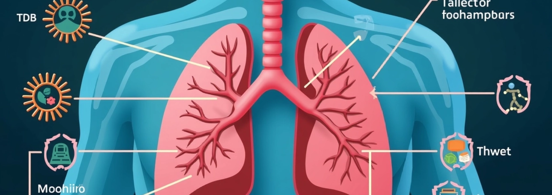
The pathophysiology of latent tuberculosis infection begins when a person inhales airborne droplets containing Mycobacterium tuberculosis bacilli. These microscopic organisms travel through the respiratory tract and eventually reach the alveolar spaces of the lungs, where they encounter the body’s first line of immune defence. The initial interaction between the bacteria and pulmonary macrophages determines whether the infection will progress to active disease or remain in a dormant state.
Bacterial load characteristics in latent TB infection
In latent tuberculosis infection, the bacterial load remains remarkably low compared to active disease. While active TB cases may harbour millions of bacilli, individuals with LTBI typically contain fewer than 10,000 organisms distributed throughout various tissues. This minimal bacterial burden explains why conventional diagnostic methods often struggle to detect latent infections. The bacteria exist in a metabolically inactive state, reducing their oxygen consumption and cellular division rates significantly. This dormancy allows them to survive in hostile environments within the host’s tissues for extended periods, sometimes decades, without causing clinical symptoms.
Granuloma formation and immune system containment
The hallmark of latent tuberculosis infection is the formation of granulomas, which serve as the immune system’s containment strategy. Within 2-8 weeks of initial infection, specialised immune cells called macrophages surround and engulf the tubercle bacilli. These cells then recruit additional immune components, including lymphocytes, epithelioid cells, and multinucleated giant cells, forming organised structures known as granulomas. The granuloma acts as a microscopic prison, preventing bacterial dissemination while maintaining the organisms in a dormant state. T-helper cells play a crucial role in maintaining granuloma integrity by secreting interferon-gamma and other cytokines that activate macrophages and sustain the inflammatory response.
Caseous necrosis and calcification processes
Over time, the central portion of granulomas may undergo caseous necrosis, creating a cheese-like material that provides a low-oxygen environment where tubercle bacilli can persist. This necrotic centre becomes surrounded by a fibrous capsule that further isolates the bacteria from the immune system. In some cases, calcium deposits form within these lesions, creating calcified granulomas visible on chest radiographs. These calcified lesions, often called Ghon complexes when they involve both lung tissue and regional lymph nodes, represent healed primary tuberculosis infections and may contain viable bacteria capable of reactivation under appropriate conditions.
Host-pathogen equilibrium in asymptomatic carriers
The maintenance of latent tuberculosis infection depends on a delicate balance between the host’s immune response and the pathogen’s survival mechanisms. Mycobacterium tuberculosis has evolved sophisticated strategies to evade immune destruction, including the ability to resist phagosome-lysosome fusion within macrophages and to modulate host cell death pathways. This equilibrium can persist for decades without clinical manifestation, but various factors can tip the balance toward bacterial reactivation. Immunosuppression, malnutrition, advanced age, and certain medical conditions can weaken the immune containment, allowing dormant bacteria to multiply and potentially progress to active tuberculosis disease.
Tuberculin skin test and Interferon-Gamma release assays for LTBI detection
Accurate diagnosis of latent tuberculosis infection relies heavily on immunological testing methods that detect the body’s cellular immune response to tubercle bacilli. The two primary diagnostic approaches include the traditional tuberculin skin test and the newer interferon-gamma release assays. Both methods have distinct advantages and limitations, making the choice of testing method dependent on patient characteristics, clinical circumstances, and available resources.
TST administration technique and mantoux method interpretation
The tuberculin skin test, also known as the Mantoux test, involves the intradermal injection of purified protein derivative (PPD) tuberculin into the volar surface of the forearm. A healthcare professional must inject exactly 0.1 mL of PPD solution containing 5 tuberculin units using a 27-gauge needle. The injection should create a pale wheal of 6-10 mm in diameter immediately after administration. Reading the test requires examination of the injection site 48-72 hours post-administration by a trained healthcare worker who measures the diameter of induration, not erythema. The interpretation of TST results depends on patient risk factors, with cut-off points of 5 mm, 10 mm, or 15 mm of induration considered positive depending on the individual’s epidemiological risk profile and immune status.
Quantiferon-gold plus laboratory analysis
The QuantiFERON-Gold Plus test represents a significant advancement in LTBI diagnostics, measuring interferon-gamma release from sensitised T-lymphocytes following stimulation with specific M. tuberculosis antigens. This blood-based assay requires a single patient visit and eliminates the subjectivity inherent in TST reading. The test uses two antigen tubes (TB1 and TB2) containing peptides from ESAT-6, CFP-10, and TB7.7 proteins, along with positive and negative control tubes. Laboratory technicians incubate whole blood samples with these antigens for 16-24 hours at 37°C, then measure interferon-gamma concentrations using enzyme-linked immunosorbent assay techniques. Results are reported as positive, negative, or indeterminate based on specific cut-off values and quality control criteria.
T-SPOT.TB Enzyme-Linked immunosorbent spot assay
The T-SPOT.TB test offers another interferon-gamma release assay option that directly enumerates interferon-gamma-producing T-cells rather than measuring cytokine concentrations. This method requires peripheral blood mononuclear cell separation and standardisation to 250,000 cells per well before stimulation with ESAT-6 and CFP-10 antigens. The enzyme-linked immunosorbent spot technique captures and visualises individual cytokine-secreting cells as distinct spots, which are then counted using automated or manual methods. This approach may provide greater sensitivity in immunocompromised patients with low lymphocyte counts, as it accounts for the actual number of responding cells rather than total cytokine production.
Cross-reactivity issues with BCG vaccination and NTM exposure
One significant challenge in LTBI diagnosis involves distinguishing true M. tuberculosis infection from cross-reactive responses caused by BCG vaccination or non-tuberculous mycobacterial (NTM) exposure. The tuberculin skin test exhibits substantial cross-reactivity with BCG vaccination, particularly when administered in infancy or repeatedly. This cross-reactivity can persist for years, leading to false-positive results that complicate LTBI diagnosis in BCG-vaccinated populations. Interferon-gamma release assays demonstrate superior specificity in this context, as they utilise antigens absent from BCG and most NTM species. However, some cross-reactivity may still occur with certain environmental mycobacteria, particularly M. kansasii and M. marinum , which share some antigenic components with M. tuberculosis .
Radiological manifestations and chest X-Ray findings in latent tuberculosis
Chest radiography plays a fundamental role in evaluating individuals suspected of having latent tuberculosis infection, primarily to exclude active pulmonary tuberculosis before initiating treatment. By definition, patients with latent TB infection should have normal chest radiographs or show only evidence of healed tuberculosis without signs of active disease. However, understanding the spectrum of radiological findings associated with previous TB exposure helps clinicians interpret imaging studies appropriately and make informed treatment decisions.
The classic radiological manifestations of healed tuberculosis include calcified granulomas, particularly in the upper lobes or superior segments of lower lobes, often accompanied by calcified hilar or mediastinal lymph nodes forming Ghon complexes. These calcifications represent the body’s successful containment of primary tuberculosis infection and may contain dormant bacilli capable of reactivation. Fibrotic scarring in the upper lung zones, particularly the apical and subapical regions, may also indicate previous tuberculosis exposure. Such scarring can appear as linear densities, volume loss, or architectural distortion without active inflammatory changes.
Healthcare professionals must carefully distinguish between inactive radiological changes and findings suggestive of active tuberculosis. Active disease typically presents with consolidation, cavitation, pleural effusions, or progressive changes on serial imaging. The presence of cavitary lesions, in particular, raises significant concern for active tuberculosis and requires thorough clinical evaluation, including sputum examination for acid-fast bacilli and mycobacterial cultures. Additionally, any radiographical abnormality accompanied by compatible clinical symptoms warrants investigation for active tuberculosis before considering LTBI treatment.
The identification and treatment of latent tuberculosis infection substantially reduces the risk that LTBI will progress to active TB disease, with treatment effectiveness reaching approximately 90% when completed appropriately.
Risk stratification and progression factors to active disease
Risk stratification forms the cornerstone of effective latent tuberculosis infection management, as not all individuals with LTBI require treatment. Healthcare providers must carefully evaluate multiple factors that influence the likelihood of progression from latent infection to active tuberculosis disease. This assessment guides treatment decisions and helps optimise resource allocation while maximising public health benefits.
Immunocompromised populations and HIV co-infection statistics
Human immunodeficiency virus infection represents the most potent risk factor for tuberculosis reactivation, increasing the annual risk of progression from approximately 0.1% to 5-10%. Individuals with CD4+ T-cell counts below 200 cells/μL face particularly elevated risks, with some studies reporting progression rates exceeding 15% annually. Other immunocompromising conditions that significantly increase reactivation risk include solid organ transplantation, haematological malignancies, and prolonged corticosteroid therapy. Patients receiving anti-TNF-alpha therapies for autoimmune conditions face 2-8 fold increased risks of tuberculosis reactivation, necessitating careful screening and preventive treatment before initiating these biologics.
Age-related reactivation patterns in elderly patients
Advanced age substantially increases tuberculosis reactivation risk due to immunosenescence and accumulated comorbidities. Adults over 65 years demonstrate 2-3 fold higher progression rates compared to younger individuals, with the risk increasing progressively with age. Elderly patients often present atypical clinical manifestations that can delay diagnosis, and they frequently have multiple comorbidities that complicate treatment decisions. The presence of diabetes mellitus, chronic kidney disease, or malnutrition further amplifies age-related risks, creating a cumulative effect that substantially increases the likelihood of progression to active tuberculosis.
Diabetes mellitus and TNF-Alpha inhibitor therapy complications
Diabetes mellitus represents an increasingly recognised risk factor for tuberculosis reactivation, particularly when poorly controlled with HbA1c levels exceeding 7%. Diabetic patients demonstrate 2-3 fold increased risks of progression to active tuberculosis, with higher risks associated with insulin dependence and diabetic complications. The mechanisms underlying this increased susceptibility include impaired cellular immunity, altered cytokine responses, and compromised macrophage function. TNF-alpha inhibitor therapies used for rheumatoid arthritis, inflammatory bowel disease, and other autoimmune conditions create profound immunosuppression that dramatically increases tuberculosis risk, with highest risks observed with infliximab and adalimumab therapy.
Malnutrition and substance abuse risk multipliers
Malnutrition significantly compromises immune function and increases tuberculosis reactivation risk, particularly when body weight falls below 90% of ideal body weight. Protein-energy malnutrition impairs T-cell function, reduces interferon-gamma production, and weakens granuloma integrity. Substance abuse, including alcohol dependence and injection drug use, creates multiple risk factors through direct immunosuppressive effects, increased exposure risks, and poor healthcare adherence. Homeless individuals face particularly elevated risks due to malnutrition, substance abuse, crowded living conditions, and limited healthcare access, creating a constellation of factors that substantially increases tuberculosis transmission and reactivation rates.
Treatment protocols and isoniazid preventive therapy regimens
Treatment of latent tuberculosis infection aims to eliminate dormant bacilli and prevent progression to active tuberculosis disease. Modern treatment approaches emphasise shorter, rifamycin-based regimens that offer improved completion rates and comparable effectiveness to traditional longer courses. The selection of appropriate treatment regimens depends on patient factors, potential drug interactions, and resistance patterns in the community or source case.
The cornerstone of LTBI treatment traditionally involved nine months of daily isoniazid monotherapy, achieving approximately 90% efficacy in preventing tuberculosis reactivation when completed appropriately. However, completion rates for this prolonged regimen often fell below 60%, limiting its public health effectiveness. Newer short-course regimens have revolutionised LTBI treatment by improving adherence while maintaining high efficacy rates. The 3HP regimen, consisting of once-weekly isoniazid and rifapentine for 12 weeks under direct observation, demonstrates equivalent efficacy to nine-month isoniazid with completion rates exceeding 80%.
Four-month daily rifampin therapy represents another effective alternative, particularly suitable for patients who cannot tolerate isoniazid or when isoniazid resistance is suspected. This regimen offers excellent completion rates and minimal side effects, making it increasingly popular among clinicians and patients. The three-month combination of daily isoniazid and rifampin provides another short-course option with high efficacy and good tolerability. Treatment selection must consider drug-susceptibility patterns of the presumed source case, with modifications necessary when drug resistance is suspected or confirmed.
Treatment for latent TB infection is 90% effective in preventing TB disease when completed appropriately, highlighting the critical importance of treatment completion and patient adherence.
Monitoring during LTBI treatment focuses primarily on detecting adverse reactions rather than assessing treatment response, as no reliable markers exist for monitoring treatment efficacy. Baseline laboratory testing typically includes liver function tests, complete blood count, and HIV testing when indicated. Monthly clinical assessments help identify potential adverse reactions, particularly hepatotoxicity associated with isoniazid and rifamycin therapy. Patient education regarding symptoms of drug toxicity, including nausea, vomiting, abdominal pain, jaundice, and unexplained fatigue, ensures early recognition and prompt medical attention when necessary.
Public health surveillance and contact tracing methodologies
Effective public health surveillance for latent tuberculosis infection requires systematic approaches to identify, test, and treat individuals at highest risk of infection and disease progression. Contact tracing represents a fundamental component of tuberculosis control programmes, focusing resources on individuals with recent exposure to infectious tuberculosis cases. This targeted approach maximises the yield of LTBI detection while optimising resource utilisation in public health settings.
Modern contact investigation protocols emphasise rapid identification and evaluation of contacts within concentric circles of exposure intensity. Close contacts, including household members, intimate partners, and individuals sharing confined air spaces with infectious cases, receive highest priority for evaluation. The investigation extends to casual contacts in workplace, school, or social settings based on factors such as infectiousness of the index case, duration of exposure, and environmental conditions. Molecular epidemiology techniques using DNA fingerprinting help confirm transmission links and identify previously unrecognised transmission chains within communities.
Data collection and reporting systems for LTBI surveillance vary considerably among jurisdictions, with some regions implementing comprehensive reporting requirements while others focus exclusively on active tuberculosis cases. Standardised performance indicators help evaluate programme effectiveness, including metrics such as contact identification rates, testing completion rates, and treatment initiation and completion rates among eligible contacts. These surveillance data inform resource allocation decisions and help identify populations and settings requiring enhanced tuberculosis control efforts.
The integration of LTBI management within broader tuberculosis elimination strategies requires coordination between clinical
services and public health departments at local, regional, and national levels. Electronic surveillance systems facilitate real-time data sharing and enable rapid response to tuberculosis outbreaks. Quality assurance programmes ensure standardised testing procedures and accurate result interpretation across different healthcare settings. Training programmes for healthcare workers emphasise the importance of LTBI screening in high-risk populations and promote evidence-based treatment approaches.
Contact tracing effectiveness depends heavily on the timeliness of index case identification and the thoroughness of exposure assessment. Digital contact tracing tools and geographic information systems increasingly support traditional epidemiological investigations by mapping potential exposure sites and identifying previously unknown contacts. Risk-based testing strategies prioritise individuals with the highest probability of infection and progression to active disease, ensuring optimal use of limited public health resources.
Performance monitoring for LTBI programmes requires robust data collection systems that track key indicators throughout the care cascade. These metrics include the proportion of identified contacts who complete testing, receive positive results, initiate treatment when indicated, and successfully complete prescribed regimens. Regular programme evaluation helps identify barriers to testing and treatment completion, informing quality improvement initiatives and policy modifications.
The success of tuberculosis elimination efforts increasingly relies on comprehensive LTBI management strategies that combine targeted testing with effective treatment programmes. International collaboration and data sharing enhance surveillance capabilities and support evidence-based policy development. As healthcare systems continue to evolve, innovative approaches to LTBI detection and treatment will play crucial roles in achieving the global goal of tuberculosis elimination by 2035.
Effective LTBI management requires a comprehensive approach that combines accurate diagnosis, appropriate risk assessment, evidence-based treatment regimens, and robust public health surveillance to maximise the impact on tuberculosis elimination efforts.