The frustrating cycle of hives appearing, fading away, and returning with seemingly no rhyme or reason affects millions of people worldwide. This perplexing skin condition, medically known as urticaria, can transform from a minor inconvenience into a chronic health challenge that significantly impacts daily life. Understanding why hives exhibit this cyclical pattern requires delving into the complex interplay of immune system responses, environmental triggers, and underlying medical conditions that drive these unpredictable outbreaks.
Chronic spontaneous urticaria, affecting approximately 1-3% of the global population, represents one of dermatology’s most challenging conditions to manage effectively. The recurring nature of these raised, itchy welts stems from multiple interconnected factors ranging from autoimmune processes to environmental sensitivities. What makes this condition particularly vexing is that in roughly 80% of cases, healthcare providers cannot identify a specific trigger, leaving patients feeling helpless as their symptoms ebb and flow unpredictably.
Immunological mechanisms behind chronic spontaneous urticaria recurrence
The underlying immunological processes driving chronic hive recurrence involve a sophisticated cascade of cellular and molecular events that can persist for months or even years. At the heart of this process lies an overactive immune response that treats harmless substances as threats, triggering inflammatory reactions that manifest as the characteristic raised, itchy wheals on the skin.
Research has revealed that chronic spontaneous urticaria often stems from autoimmune dysfunction, where the body’s immune system mistakenly attacks its own tissues. Approximately 50% of patients with chronic hives have detectable autoantibodies that target either the high-affinity IgE receptor or IgE antibodies themselves, creating a self-perpetuating cycle of inflammation.
Mast cell degranulation and histamine release pathways
Mast cells, the primary effector cells in urticaria development, function as immune system sentinels positioned strategically beneath the skin’s surface. When activated, these cells undergo degranulation, releasing a potent cocktail of inflammatory mediators including histamine, leukotrienes, and prostaglandins. This process explains why hives can appear and disappear so rapidly—histamine’s effects typically peak within 15-30 minutes and gradually subside over several hours.
The cyclical nature of hive recurrence often reflects the ongoing sensitisation of mast cell populations in affected skin areas. Even after visible symptoms resolve, these cells may remain in a heightened state of reactivity, primed to respond to minimal stimuli that wouldn’t affect normal skin. This phenomenon, known as priming , can persist for weeks or months, explaining why patients experience repeated outbreaks in similar body regions.
Ige-mediated type I hypersensitivity reactions in hive formation
Type I hypersensitivity reactions represent the classic allergic pathway leading to immediate hive formation. When allergens cross-link bound IgE antibodies on mast cell surfaces, they trigger rapid degranulation and symptom onset within minutes. However, in chronic spontaneous urticaria, this pathway becomes dysregulated, with elevated baseline IgE levels and inappropriate responses to normally harmless stimuli.
The persistence of elevated IgE levels in chronic urticaria patients creates a state of ongoing immune vigilance. Studies indicate that patients with chronic hives often have total IgE levels 2-3 times higher than healthy individuals, contributing to their increased reactivity. This elevated immune baseline means that even minor exposures to potential allergens can trigger disproportionate responses, leading to the characteristic unpredictable pattern of symptom recurrence.
Complement system activation and C3a/C5a anaphylatoxin role
The complement system, a crucial component of innate immunity, plays a significant role in chronic urticaria through the generation of anaphylatoxins C3a and C5a. These powerful inflammatory mediators directly stimulate mast cell degranulation and increase vascular permeability, contributing to the formation of wheals and the surrounding inflammatory response.
Complement activation can occur through multiple pathways in chronic urticaria, including direct activation by autoantibodies or through immune complex formation. Research has shown that C3a and C5a levels remain elevated in many chronic urticaria patients even during symptom-free periods, suggesting ongoing subclinical inflammation. This persistent complement activation may explain why some patients experience sudden symptom flares without obvious triggers—the inflammatory machinery remains constantly primed for activation.
Autoimmune thyroid disease association with chronic urticaria
The strong association between autoimmune thyroid disease and chronic urticaria affects approximately 20-30% of chronic hive sufferers. This connection extends beyond mere coincidence, reflecting shared autoimmune mechanisms and genetic predispositions. Patients with Hashimoto’s thyroiditis or Graves’ disease demonstrate significantly higher rates of chronic urticaria, often experiencing more severe and treatment-resistant symptoms.
Thyroid dysfunction can influence hive recurrence through multiple mechanisms, including alterations in immune function, changes in skin sensitivity, and modifications in drug metabolism. Even patients with subclinical thyroid dysfunction—those with normal hormone levels but positive thyroid antibodies—show increased susceptibility to chronic urticaria. This relationship underscores the importance of comprehensive thyroid evaluation in patients with recurrent hives, as optimising thyroid function often leads to significant improvement in skin symptoms.
Environmental and dietary triggers causing intermittent hive manifestation
Environmental and dietary factors represent some of the most identifiable yet variable triggers for hive recurrence. Unlike the constant immune dysfunction underlying chronic spontaneous urticaria, these external factors create intermittent exposure patterns that directly correlate with symptom flares. Understanding these triggers becomes crucial for patients seeking to gain some measure of control over their unpredictable symptoms.
The challenge with environmental triggers lies in their ubiquity and the delayed reactions they can sometimes provoke. What appears to be a random hive outbreak may actually result from cumulative exposures or delayed hypersensitivity reactions occurring hours or even days after initial contact. This temporal disconnect makes trigger identification particularly challenging for both patients and healthcare providers.
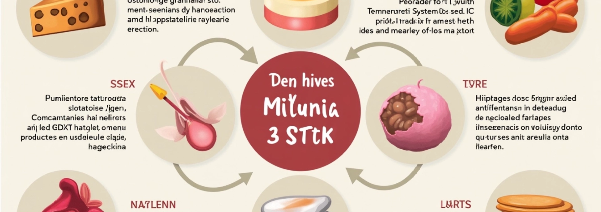
Histamine-rich foods: aged cheese, fermented products, and wine reactions
Histamine intolerance represents a significant yet often overlooked trigger for recurrent hives. Foods naturally high in histamine or those that promote histamine release can trigger symptoms in susceptible individuals through a non-allergic pathway. Aged cheeses, fermented vegetables, cured meats, and alcoholic beverages contain particularly high histamine concentrations that can overwhelm the body’s natural histamine-degrading capacity.
The enzyme diamine oxidase (DAO) normally breaks down dietary histamine in the intestinal tract. However, genetic variations, medications, or intestinal inflammation can reduce DAO activity, leading to histamine accumulation and subsequent hive formation. This mechanism explains why some patients notice symptom patterns related to specific meals or dietary periods, particularly when consuming multiple histamine-rich foods simultaneously. Wine reactions deserve special attention, as alcohol not only contains histamine but also inhibits DAO enzyme activity, creating a double effect that can trigger severe symptoms even in individuals who normally tolerate moderate histamine intake.
Salicylate sensitivity in aspirin and natural food sources
Salicylate sensitivity affects approximately 2-23% of chronic urticaria patients and represents one of the most clinically relevant yet underdiagnosed triggers. Salicylates occur naturally in many fruits, vegetables, herbs, and spices, making complete avoidance challenging. Aspirin and other NSAIDs contain synthetic salicylates that can trigger immediate reactions, but natural dietary sources often cause more subtle, delayed responses that patients may not associate with their symptoms.
The mechanism of salicylate-induced urticaria involves cyclooxygenase-1 (COX-1) enzyme inhibition, which shifts arachidonic acid metabolism towards leukotriene production. These inflammatory mediators directly stimulate mast cell degranulation and can maintain chronic inflammation for extended periods. Foods highest in natural salicylates include berries, grapes, tomatoes, peppers, and most herbs and spices. Patients with salicylate sensitivity often report improvement with low-salicylate diets , though such restrictive approaches require careful nutritional planning to prevent deficiencies.
Temperature-dependent urticaria: Cold-Induced and Heat-Induced variants
Physical urticaria represents a distinct category where environmental temperature changes directly trigger hive formation through non-immunological mechanisms. Cold urticaria affects approximately 15% of chronic urticaria patients and can range from mild reactions to cold air or water to severe systemic responses requiring emergency treatment. The condition involves abnormal mast cell activation in response to temperature changes, with symptoms typically appearing within minutes of exposure and resolving within hours of rewarming.
Heat-induced urticaria, while less common, can be equally disruptive to daily life. This condition encompasses cholinergic urticaria triggered by sweating and exercise, as well as direct heat urticaria from contact with warm objects. The mechanisms involve either direct mast cell activation by temperature changes or indirect activation through neurogenic pathways. Cholinergic urticaria specifically involves acetylcholine-mediated mast cell degranulation, explaining why emotional stress, spicy foods, and physical exercise can all trigger similar symptoms. These temperature-dependent reactions often follow seasonal patterns, with cold urticaria peaking in winter months and heat-related variants becoming more problematic during summer.
Contact allergens: latex, nickel, and Fragrance-Induced episodes
Contact allergens represent immediate environmental triggers that can cause localised hive formation at contact sites, though systemic reactions are also possible in highly sensitised individuals. Latex sensitivity has become increasingly recognised as a cause of both occupational and household-related hive outbreaks, affecting healthcare workers, food service employees, and individuals with frequent medical device exposure.
Nickel allergy, one of the most common contact sensitivities worldwide, can trigger hives through direct skin contact with jewellery, clothing fasteners, coins, and electronic devices. The reaction typically begins at contact sites but can generalise in severely allergic individuals. Fragrance sensitivity represents a complex trigger involving hundreds of potential chemical compounds found in personal care products, cleaning supplies, and environmental sources. These reactions can occur through direct skin contact, inhalation, or even indirect exposure through contaminated surfaces. The challenge with contact allergens lies in their omnipresence in modern environments and the difficulty of achieving complete avoidance without significant lifestyle modifications.
Pharmacological factors influencing urticaria persistence and resolution
Medications represent both potential triggers for hive development and essential tools for symptom management, creating a complex relationship that significantly influences the condition’s course. Understanding these pharmacological interactions becomes crucial for optimising treatment outcomes while avoiding drug-induced complications that can perpetuate or worsen symptoms.
The paradox of medication-induced urticaria lies in the fact that drugs intended to treat other conditions can sometimes trigger or maintain the very symptoms patients seek to control. This situation requires careful balancing of therapeutic benefits against potential adverse effects, often necessitating medication adjustments or alternative treatment approaches.
ACE Inhibitor-Induced angioedema and urticaria patterns
Angiotensin-converting enzyme (ACE) inhibitors represent one of the most clinically significant medication classes associated with delayed-onset urticaria and angioedema. Unlike immediate allergic reactions, ACE inhibitor-induced symptoms can develop weeks, months, or even years after starting treatment, making the connection difficult to establish. This delayed onset occurs because ACE inhibitors interfere with bradykinin metabolism, leading to accumulation of this potent vasodilator and inflammatory mediator.
The bradykinin pathway operates independently of histamine-mediated mechanisms, explaining why ACE inhibitor-induced reactions often resist standard antihistamine treatment. Patients may experience recurrent episodes of lip, tongue, or facial swelling accompanied by urticarial lesions, particularly during periods of physiological stress when bradykinin levels naturally increase. African American patients show higher susceptibility to ACE inhibitor-induced angioedema, with incidence rates 3-5 times higher than other populations. Recognition of this pattern is crucial because continued ACE inhibitor use can lead to life-threatening airway obstruction, requiring immediate drug discontinuation and alternative antihypertensive therapy.
Nsaid-exacerbated respiratory disease and skin manifestations
Non-steroidal anti-inflammatory drugs (NSAIDs) can trigger urticaria through multiple pathways, with aspirin-exacerbated respiratory disease (AERD) representing the most severe manifestation. This condition combines chronic rhinosinusitis, nasal polyps, asthma, and cutaneous reactions including chronic urticaria. The underlying mechanism involves COX-1 enzyme inhibition, which redirects arachidonic acid metabolism towards increased leukotriene production.
NSAID-induced urticaria can manifest as acute reactions occurring within hours of ingestion or as chronic symptoms maintained by regular NSAID use. Cross-reactivity between different NSAIDs is common, though selective COX-2 inhibitors may be better tolerated in some patients. The condition affects approximately 10-20% of chronic urticaria patients, making NSAID avoidance a crucial component of comprehensive management. Aspirin desensitisation protocols exist for patients requiring ongoing anti-inflammatory therapy, though this approach requires specialised medical supervision and careful monitoring for severe reactions.
Antihistamine tolerance development and H1 receptor desensitisation
Long-term antihistamine therapy, while generally safe and effective, can sometimes lead to tolerance development through H1 receptor desensitisation or upregulation of alternative inflammatory pathways. Patients may notice diminishing effectiveness of previously successful treatments, requiring dose escalations or medication changes to maintain symptom control. This phenomenon appears more common with first-generation antihistamines due to their broader receptor activity and shorter duration of action.
Second-generation antihistamines like cetirizine, loratadine, and fexofenadine demonstrate more consistent long-term efficacy due to their selective H1 receptor binding and longer half-lives. However, even these medications can show reduced effectiveness over time in some patients. The mechanism likely involves adaptive changes in mast cell sensitivity and alternative degranulation pathways that bypass H1 receptor blockade. Current treatment guidelines recommend increasing antihistamine doses up to four times the standard amount before considering treatment failure, as many patients achieve excellent control with higher doses rather than requiring alternative medications.
Corticosteroid withdrawal effects on chronic urticaria management
Systemic corticosteroids provide rapid and dramatic improvement in acute urticaria flares but can create problematic rebound effects when discontinued. The anti-inflammatory potency of corticosteroids suppresses virtually all aspects of the allergic and inflammatory response, often leading to complete symptom resolution within 24-48 hours. However, this powerful suppression can be followed by severe symptom rebound when medications are tapered too quickly or discontinued abruptly.
Corticosteroid rebound typically occurs 3-7 days after discontinuation and can manifest as symptoms more severe than the original presentation. This phenomenon results from the sudden loss of anti-inflammatory suppression combined with the time required for natural regulatory mechanisms to re-establish homeostasis. Long-term corticosteroid use in chronic urticaria is generally discouraged due to significant adverse effects, but short courses remain valuable for managing severe flares. Proper tapering schedules and bridging with alternative anti-inflammatory treatments can help minimise rebound effects while maintaining symptom control during the transition period.
Stress-related psychoneuroimmunology in hive cyclical patterns
The intricate relationship between psychological stress and urticaria recurrence represents one of the most fascinating yet challenging aspects of chronic hive management. Stress-induced symptoms occur through well-established psychoneuroimmunological pathways that directly influence immune function and inflammatory responses. Understanding these connections provides insight into why many patients notice strong correlations between their emotional state and symptom severity.
Chronic stress creates a state of immune dysregulation that mirrors many aspects of autoimmune dysfunction seen in spontaneous urticaria. The hypothalamic-pituitary-adrenal (HPA) axis becomes hyperactive during stress periods, leading to elevated cortisol levels that initially suppress inflammation but eventually lead to immune rebound effects when stress subsides. This cyclical pattern can create predictable hive outbreaks following stressful periods, even when obvious environmental triggers are absent.
Research demonstrates that psychological stress directly affects mast cell function through neural pathways connecting the brain to peripheral immune cells. The neuropeptide substance P, released during stress responses, can directly trigger mast cell degranulation independent of allergic pathways. Additionally, stress hormones alter the production and effectiveness of natural antihistamines and anti-inflammatory compounds, reducing the body’s ability to control inflammatory responses.
The psychological burden of chronic urticaria extends beyond mere physical discomfort, creating anxiety and depression that can perpetuate the inflammatory cycle. Many patients develop anticipatory anxiety about potential outbreaks, which itself triggers stress hormones that increase the likelihood of symptoms. This creates a self-reinforcing pattern where fear of hives increases the probability of their occurrence, establishing a psychological component that must be addressed for successful long-term management.
Underlying medical conditions masquerading as idiopathic urticaria
Many cases initially diagnosed as chronic idiopathic urticaria actually stem from unrecognised underlying medical conditions that drive persistent inflammation and immune dysfunction. These masked conditions can perpetuate hive symptoms for years if left undiagnosed, highlighting the importance of comprehensive medical evaluation beyond basic allergy testing. The interconnected nature of immune system dysfunction means that seemingly unrelated conditions can manifest as chronic skin symptoms.
Chronic infections represent one of the most overlooked categories of underlying triggers. Helicobacter pylori gastric infections affect approximately 15-20% of chronic urticaria patients and can maintain inflammatory responses through molecular mimicry and chronic antigen exposure. Similarly, dental infections, chronic sinusitis, and parasitic infections can provide ongoing antigenic stimulation that perpetuates mast cell activation and histamine release.
Autoimmune connective tissue disorders, particularly systemic lupus erythematosus and Sjögren’s syndrome, frequently present with urticarial lesions as early manifestations before classic disease features develop. These conditions create systemic inflammation and immune complex formation that can trigger chronic hives months or years before other diagnostic criteria are met. Complement consumption and autoantibody production in these disorders directly activate the same pathways involved in spontaneous urticaria.
Malignancy represents a rare but important consideration in chronic urticaria evaluation, particularly in older patients or those with treatment-resistant symptoms. Haematological malignancies, including lymphomas and leukaemias, can present with paraneoplastic urticaria through immune system dysregulation and abnormal cytokine production. Solid organ tumours may also trigger chronic inflammation and immune responses that manifest as persistent skin symptoms.
Diagnostic approaches for identifying recurrent hive triggers
Effective diagnosis of recurrent hive triggers requires a systematic approach combining detailed history-taking, targeted testing, and elimination strategies. The complexity of potential triggers means that identifying specific causes often requires patience and methodical investigation over several months. However, this investment in comprehensive evaluation frequently yields significant improvements in symptom control and quality of life.
Patient symptom diaries represent the cornerstone of trigger identification, providing objective data about symptom patterns, timing, and potential associations. Detailed logs should document food intake, medications, activities, stress levels, weather conditions, and symptom severity using standardised scales. Digital applications and wearable devices can enhance data collection accuracy and identify subtle patterns that might otherwise be missed. Analysis of diary data over 4-6 weeks often reveals correlations that guide targeted testing and elimination trials.
Laboratory evaluation should include comprehensive assessment of inflammatory markers, immune function, and potential underlying conditions. Complete blood count with differential can identify eosinophilia suggesting parasitic infections or drug reactions, while elevated ESR or CRP levels indicate systemic inflammation requiring further investigation. Thyroid function tests, including TSH, free T4, and thyroid peroxidase antibodies, are essential given the strong association between thyroid autoimmunity and chronic urticaria.
Complement levels (C3, C4, CH50) provide insight into immune system activation and consumption, while tryptase levels can identify underlying mastocytosis or increased mast cell burden. Specific IgE testing should focus on suspected allergens identified through history and diary analysis rather than broad panels that may yield irrelevant positive results. Autologous serum skin testing can identify patients with functional autoantibodies, though this test requires specialised interpretation and may not be widely available.
Elimination diets represent powerful diagnostic tools when properly implemented with professional guidance. The most effective approach involves initial restriction to a limited number of low-allergenic foods, followed by systematic reintroduction of suspected triggers while monitoring symptoms. Histamine-restricted diets can identify histamine intolerance, while salicylate-free diets help diagnose salicylate sensitivity. These elimination protocols require 2-4 weeks for initial restriction and several additional weeks for systematic reintroduction, demanding significant patient commitment and nutritional oversight.
Physical provocation testing can diagnose specific forms of physical urticaria through controlled exposure to relevant stimuli. Ice cube testing for cold urticaria, exercise challenge for cholinergic urticaria, and dermographism assessment through controlled skin stroking provide objective diagnostic information. These tests should be performed in controlled medical settings with appropriate emergency management capabilities, as severe reactions can occasionally occur.
Advanced diagnostic approaches may include complement functional assays, flow cytometry for immune cell populations, and specialised autoantibody testing for rare forms of autoimmune urticaria. Patch testing with extended allergen panels can identify delayed-type hypersensitivity reactions, while histamine release assays provide functional assessment of mast cell reactivity. These sophisticated tests typically require referral to specialised centres and may be reserved for particularly challenging cases resistant to standard evaluation and treatment approaches.