Revolutionary advances in neuroimaging technology have transformed our understanding of how individual brain patterns influence behaviour, cognition, and mental health. Dr. Daniel Amen’s comprehensive brain assessment protocol combines cutting-edge functional brain imaging with detailed neuropsychiatric evaluation to identify specific brain patterns and optimise mental performance. This innovative approach has analysed over 200,000 brain scans from patients across 155 countries, revealing distinct neurological signatures that correlate with various psychological and cognitive conditions.
The assessment methodology extends far beyond traditional psychiatric evaluation, incorporating advanced neuroimaging techniques that visualise brain activity in real-time. Unlike conventional approaches that rely solely on symptom observation, this comprehensive system examines actual brain function to identify underlying neurological patterns. The protocol has revolutionised treatment approaches for attention deficit disorders, depression, anxiety, and traumatic brain injuries by providing objective, measurable data about brain performance.
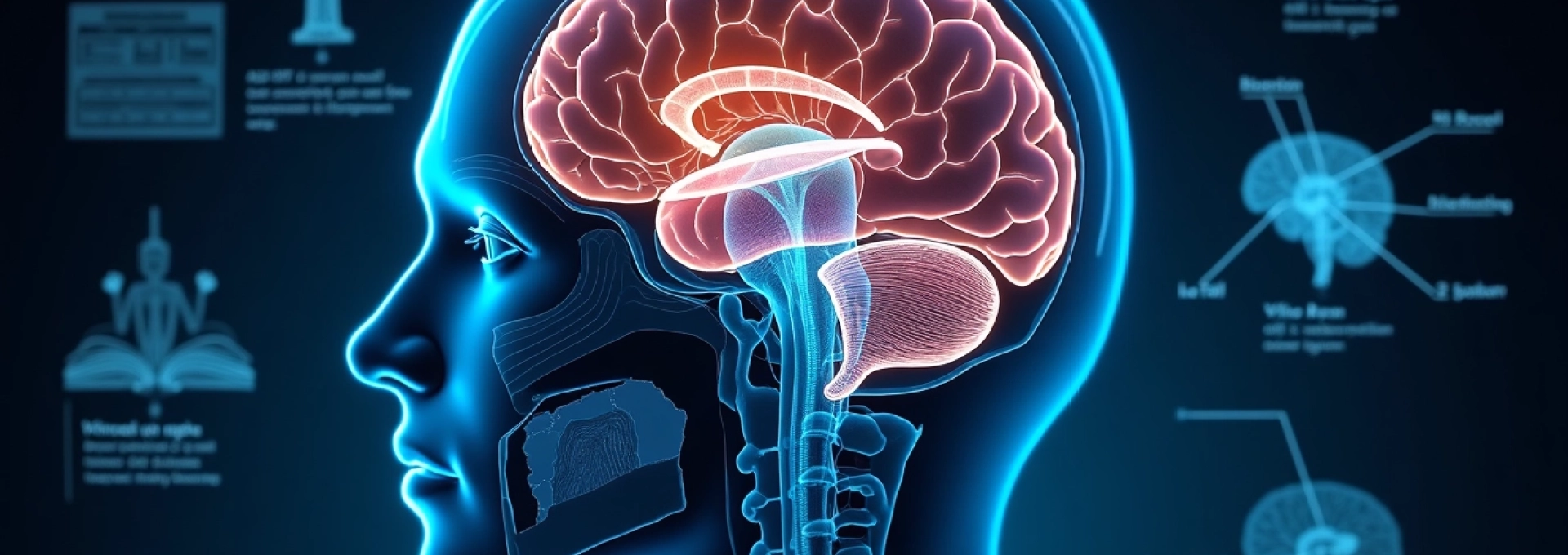
SPECT brain imaging technology in dr. amen’s assessment protocol
Single Photon Emission Computed Tomography (SPECT) imaging forms the cornerstone of Dr. Amen’s neuropsychiatric evaluation system. This sophisticated nuclear medicine technique captures detailed images of brain blood flow and metabolic activity, providing unprecedented insights into functional brain patterns that remain invisible to conventional diagnostic methods. The technology enables clinicians to observe how different brain regions communicate and function during various cognitive tasks and emotional states.
Single photon emission computed tomography methodology
The SPECT imaging process involves injecting a small amount of radioactive tracer into the bloodstream, which travels to the brain and binds to areas of high metabolic activity. Specialised gamma cameras then rotate around the patient’s head, capturing multiple images from different angles to create three-dimensional representations of brain function. This non-invasive procedure typically takes 30-45 minutes and provides detailed visualisation of cerebral blood flow patterns across all brain regions.
The resulting images reveal areas of normal, increased, or decreased brain activity with remarkable precision. Regions showing optimal blood flow appear as bright areas on the scan, while areas with reduced activity appear darker. This functional mapping capability allows clinicians to identify specific brain regions that may be contributing to symptoms such as inattention, mood instability, or cognitive difficulties.
Cerebral blood flow pattern analysis
Cerebral blood flow patterns provide crucial information about brain health and functionality, as blood flow directly correlates with neural activity and metabolic demands. The SPECT analysis examines flow patterns across multiple brain regions, including the prefrontal cortex, anterior cingulate gyrus, temporal lobes, limbic system, and basal ganglia. Each region’s activity level is compared to established normative databases to identify areas of dysfunction.
The analysis reveals distinct patterns associated with various neuropsychiatric conditions. For instance, decreased prefrontal cortex activity often correlates with attention difficulties and impulse control problems, while hyperactivity in the anterior cingulate gyrus may indicate obsessive-compulsive tendencies or cognitive inflexibility. These patterns provide objective evidence for tailored treatment approaches targeting specific brain regions.
Technetium-99m HMPAO radiotracer application
The radiotracer Technetium-99m HMPAO (hexamethylpropyleneamine oxime) serves as the primary imaging agent for SPECT brain scans. This compound crosses the blood-brain barrier efficiently and becomes trapped in brain tissue proportional to regional blood flow at the time of injection. The tracer’s unique properties allow for accurate measurement of cerebral perfusion patterns without affecting normal brain function.
The radiotracer has a half-life of approximately six hours, ensuring adequate imaging time while minimising radiation exposure. The amount of radiation received during a SPECT scan is comparable to that of a chest X-ray, making it a safe diagnostic tool for most patients. The tracer’s distribution remains stable for several hours after injection, allowing for detailed image acquisition without time pressure.
Functional neuroimaging vs structural MRI comparison
While structural MRI provides excellent anatomical detail of brain structure, SPECT imaging offers complementary functional information about brain activity patterns. MRI excels at detecting structural abnormalities such as tumours, lesions, or anatomical variations but cannot assess how well different brain regions are functioning. SPECT imaging fills this gap by revealing metabolic activity patterns that may appear normal on structural scans.
Many neuropsychiatric conditions involve functional rather than structural abnormalities, making SPECT imaging particularly valuable for diagnosis and treatment planning. The combination of both imaging modalities provides a comprehensive picture of brain health, addressing both anatomical and functional aspects of neurological conditions.
Comprehensive neuropsychiatric evaluation components
The complete brain assessment protocol incorporates multiple evaluation components beyond SPECT imaging to provide a thorough understanding of cognitive and emotional functioning. This multi-dimensional approach ensures that all aspects of brain health are examined, from basic neurological function to complex psychological patterns. The comprehensive evaluation typically requires 4-6 hours and involves various standardised tests and assessments administered by trained professionals.
Amen clinics brain system checklist assessment
The Brain System Checklist represents a sophisticated symptom assessment tool that evaluates function across six major brain systems: prefrontal cortex, anterior cingulate gyrus, deep limbic system, basal ganglia, temporal lobes, and cerebellum. Each system’s checklist contains specific questions designed to identify symptoms associated with dysfunction in that particular brain region. The assessment helps correlate clinical symptoms with anticipated SPECT imaging findings.
Patients complete detailed questionnaires addressing attention, memory, mood, anxiety, impulse control, and social behaviour patterns. The checklist includes questions about sleep patterns, energy levels, motivation, and cognitive flexibility. This systematic approach ensures that all relevant symptoms are documented and correlated with brain imaging results for comprehensive treatment planning.
Cognitive performance testing battery
Standardised cognitive testing evaluates specific aspects of brain function including attention, memory, executive function, processing speed, and visual-spatial abilities. The testing battery incorporates validated neuropsychological instruments such as continuous performance tests, memory assessments, and executive function evaluations. These tests provide objective measures of cognitive performance that complement subjective symptom reports.
The cognitive assessment typically includes computerised attention tests that measure sustained attention, impulse control, and response variability. Memory testing evaluates both short-term and long-term memory function across verbal and visual domains. Executive function tests assess planning abilities, cognitive flexibility, and problem-solving skills. Results are compared to age-matched normative data to identify specific areas of cognitive strength and weakness.
Clinical interview and psychiatric history analysis
Comprehensive clinical interviews explore detailed personal, family, and medical histories to identify factors that may influence brain function. The interview process examines developmental milestones, academic and occupational performance, relationship patterns, and previous mental health treatments. Family history assessment identifies genetic predispositions to neuropsychiatric conditions and helps inform treatment recommendations.
The psychiatric history review includes detailed examination of previous diagnoses, medications, and treatment responses. This information helps clinicians understand patterns of symptom presentation and treatment efficacy. The interview also explores lifestyle factors such as diet, exercise, sleep patterns, and substance use that may impact brain health and treatment outcomes.
Quantitative EEG brainwave pattern evaluation
Quantitative electroencephalography (qEEG) measures electrical activity across the brain’s surface, providing information about brainwave patterns and cortical function. The assessment records brain activity from multiple electrode sites and compares patterns to normative databases. qEEG can identify abnormal brainwave patterns associated with attention disorders, mood disturbances, and cognitive dysfunction.
The qEEG evaluation measures different brainwave frequencies including delta, theta, alpha, and beta waves. Abnormal patterns may indicate areas of brain dysfunction or inefficiency. For example, excessive theta activity in frontal regions often correlates with attention difficulties, while abnormal alpha patterns may suggest mood regulation problems. The qEEG data provides additional objective information to complement SPECT imaging findings.
Brain pattern type classification system
Dr. Amen’s research has identified 16 distinct brain types based on SPECT imaging patterns and clinical presentations. These brain types represent different patterns of neural activity that influence personality traits, cognitive abilities, and susceptibility to various mental health conditions. The classification system moves beyond traditional psychiatric diagnoses to focus on underlying brain function patterns that drive behaviour and symptoms.
The five primary brain types include Balanced, Spontaneous, Persistent, Sensitive, and Cautious patterns, each with specific characteristics and treatment recommendations. The Balanced type shows optimal activity across all brain regions, while the Spontaneous type typically exhibits decreased prefrontal cortex activity. The Persistent type demonstrates increased anterior cingulate activity, the Sensitive type shows heightened deep limbic activity, and the Cautious type displays increased basal ganglia activity.
Each brain type has associated strengths and vulnerabilities that influence daily functioning and mental health risk factors. Understanding your specific brain type enables targeted interventions that work with your brain’s natural patterns rather than against them. The classification system provides personalised recommendations for nutrition, supplements, exercise, and lifestyle modifications based on individual brain patterns.
The remaining 11 combination brain types represent various combinations of the primary patterns, reflecting the complexity of human brain function. These combination types help explain why individuals may exhibit mixed symptoms or respond differently to similar treatments. The comprehensive typing system ensures that treatment recommendations are tailored to each person’s unique neurobiological profile.
ADD/ADHD subtype identification through brain imaging
Traditional ADHD diagnosis relies primarily on behavioural observations and symptom checklists, often leading to one-size-fits-all treatment approaches. Dr. Amen’s brain imaging research has identified seven distinct ADHD subtypes, each with unique brain patterns and optimal treatment strategies. This neurobiologically-based approach has revolutionised ADHD treatment by matching interventions to specific brain dysfunction patterns rather than general symptom presentations.
Classic ADD prefrontal cortex hypoactivity
Classic ADD presents with decreased activity in the prefrontal cortex, particularly during concentration tasks. This brain pattern correlates with symptoms of inattention, distractibility, disorganisation, and impulse control difficulties. SPECT scans typically show reduced blood flow to frontal brain regions, especially when patients attempt to concentrate or focus on demanding tasks.
Individuals with this subtype often benefit from stimulant medications that increase dopamine and norepinephrine activity in frontal brain regions. The hypoactive prefrontal cortex responds well to interventions that increase neural stimulation, including certain supplements, dietary modifications, and cognitive training exercises. Treatment success rates improve significantly when interventions target the specific brain dysfunction pattern rather than general ADHD symptoms.
Inattentive ADD anterior cingulate dysfunction
The Inattentive subtype combines prefrontal cortex underactivity with anterior cingulate gyrus dysfunction, resulting in attention problems without significant hyperactivity. Brain scans show decreased activity in both frontal regions and the anterior cingulate, which affects cognitive flexibility and attention switching. Patients often appear spacey or daydreamy and have difficulty following conversations or instructions.
This subtype requires treatment approaches that address both attention and cognitive flexibility issues. Stimulant medications may help with attention, but additional interventions targeting serotonin function are often necessary to address anterior cingulate dysfunction. Cognitive-behavioural therapy and mindfulness training can be particularly beneficial for improving attention regulation and mental flexibility.
Over-focused ADD anterior cingulate hyperactivity
Over-focused ADD presents with normal or decreased prefrontal activity combined with excessive anterior cingulate activity. This pattern creates attention problems accompanied by obsessive thoughts, compulsive behaviours, and difficulty shifting attention between tasks. SPECT scans reveal hyperactivity in the anterior cingulate gyrus, often extending into related brain regions.
Traditional stimulant medications may worsen symptoms in this subtype by further increasing anterior cingulate activity. Treatment typically involves serotonin-enhancing interventions such as SSRIs, combined with cognitive flexibility training and stress management techniques. Dietary modifications that naturally increase serotonin production can also be beneficial for this brain pattern.
Temporal lobe ADD limbic system irregularities
Temporal Lobe ADD combines attention difficulties with temporal lobe dysfunction, resulting in symptoms such as memory problems, emotional instability, and sometimes aggressive behaviour. Brain imaging reveals irregular activity patterns in temporal lobe regions, which may include areas of both increased and decreased function. This subtype often has a history of head injuries or infections affecting the temporal lobes.
Treatment approaches must address both attention deficits and temporal lobe stabilisation. Anticonvulsant medications are often more effective than stimulants for this subtype. Nutritional support for brain health, stress management, and memory training exercises form important components of the treatment plan. The complex nature of this subtype requires carefully coordinated interventions targeting multiple brain systems.
Depression and anxiety brain pattern recognition
Depression and anxiety disorders exhibit distinct brain patterns that can be visualised through SPECT imaging, enabling more targeted treatment approaches. Traditional mental health treatment often relies on trial-and-error medication selection, but brain imaging reveals specific patterns of neural dysfunction that predict treatment response. The imaging findings help clinicians select interventions that address underlying brain dysfunction rather than just managing symptoms.
Depression commonly presents with increased activity in the deep limbic system, particularly the limbic cortex and anterior cingulate gyrus. This hyperactivity correlates with negative thinking patterns, emotional sensitivity, and mood instability. Some depression subtypes also show decreased prefrontal activity, affecting motivation and executive function. The specific pattern of brain dysfunction influences treatment selection and prognosis.
Anxiety disorders typically demonstrate increased activity in the basal ganglia, amygdala, and other anxiety-processing brain regions. The heightened activity in these areas creates chronic worry, physical tension, and avoidance behaviours. Different anxiety disorders show distinct patterns, with generalised anxiety affecting different brain regions than panic disorder or social anxiety. Personalised treatment approaches based on specific brain patterns achieve better outcomes than generic anxiety treatments.
The brain imaging findings help predict medication response and identify patients who may benefit from alternative treatments. For instance, patients with certain brain patterns respond better to psychotherapy than medication, while others require specific types of medications targeting particular neurotransmitter systems. This precision medicine approach reduces treatment trial periods and improves patient outcomes.
Traumatic brain injury and toxic exposure detection
SPECT imaging excels at detecting functional brain damage from traumatic injuries and toxic exposures that may not appear on structural brain scans. Many patients with persistent cognitive and emotional symptoms following head injuries show normal CT and MRI scans, leading to dismissal of their complaints. SPECT imaging reveals areas of decreased blood flow and metabolic dysfunction that explain ongoing symptoms and guide treatment planning.
Traumatic brain injury patterns typically show areas of decreased activity in regions affected by the injury mechanism. Frontal and temporal regions are commonly affected due to their vulnerability during impact injuries. The imaging findings correlate with specific symptom patterns, helping clinicians understand the neurological basis for cognitive, emotional, and behavioural changes following injury. This objective evidence validates patient complaints and supports appropriate treatment interventions.
Toxic exposures from substances such as alcohol, drugs, environmental toxins, or medications can create distinctive brain patterns visible on SPECT scans. Chronic alcohol use typically shows decreased activity throughout the brain, with particular effects on frontal and cerebellar regions. Drug toxicity creates specific patterns depending on the substance involved. Environmental toxins such as mould exposure or heavy metals can cause widespread brain dysfunction with characteristic imaging findings.
The identification of toxic brain damage enables targeted treatment approaches focused on brain healing and restoration. Treatment protocols may include detoxification support, nutritional rehabilitation, hyperbaric oxygen therapy, and neuroprotective interventions. Regular follow-up imaging can monitor treatment progress and brain recovery over time. Early identification and treatment of toxic brain damage can prevent permanent neurological dysfunction and improve long-term outcomes for affected individuals.